Paleoanthropologists from Institute of Vertebrate Paleontology and Paleoanthropology (IVPP), Chinese Academy of Sciences, and Washington University in St. Louis, reported a neurocranial abnormality previously undescribed in Pleistocene human fossils, an enlarged parietal foramen (EPF) in the early Late Pleistocene Xujiayao 11 parietal bones from the Xujiayao (Houjiayao) site, in the Nihewan Basin of northern China, suggesting unusual population dynamics, most likely from high levels of inbreeding and local population instability. Researchers published their results March 18 in the journal PLOS ONE, and provided a background for understanding populational and cultural dynamics through much of human evolution.
The Xujiayao 11 neurocranial specimen was excavated during the 1977 field season at the Xujiayao site (Locality 74093 in the village of Houjiayao; 40°06’02’’ N, 113°58’39’’E). The site is situated on the west bank of the Liyi River, a small tributary of the Sanggan River, near the northwestern boundary of the Nihewan Basin, northern China. The Xujiayao human remains likely derive from early Late Pleistocene (MIS 5 to 4) deposits. Morphologically, they represent late archaic humans and are distinct from H. erectus and early modern humans.
Xujiayao 11 is a pair of partial posteromedial parietal bones from an adult. It exhibits thick cranial vault bones, arachnoid granulations, a deviated posterior sagittal suture, and a unilateral (right) parietal lacuna with a posteriorly-directed and enlarged endocranial vascular sulcus.
The Xujiayao 11 single perforation is situated on the posterior sagittal suture, close to the usual location of a normal parietal foramen. The edge margins of the perforation are rounded and beveled from outer table to the inner bone. It appears to have connected with the endocranial venous system, as is indicated by the posterior vascular sulcus. The lacuna is also associated with a right deviation of the sagittal suture. Morphologically, the Xujiayao 11 perforation therefore corresponds to an EPF, in terms of its form, position, and probable endocranial vascular connection.
"Differential diagnosis indicates that the perforation is a congenital defect, an enlarged parietal foramen, commonly associated with cerebral venous and cranial vault anomalies. It was not lethal given the individual’s age-at-death, but it may have been associated with secondary neurological deficiencies”, said first author Dr. WU Xiujie of the IVPP, “The Xujiayao 11 single perforation is consistent with modern humans diagnosed with a rare genetic mutation in the homeobox genes ALX4 on chromosome 11 and MSX2 on chromosome 5”.
EPF derive from a malformation of the parietal bones, in which normal symmetrical fetal openings in the parietal bones fail to close during the second half of pregnancy in about one out of every 25,000 modern human births. They can be circular, oblique or irregular in shape and occur near the sagittal suture a few centimeters anterior of the lambda, in the vicinity of the normal parietal foramina. The edges of the EPF are often smoothly beveled at the expense of the outer table with a resultant difference in the internal and external measurements of the foramina. They may be asymptomatic, but they are often associated with cerebral venous anomalies, irregular suture fusion and deviations of the sagittal suture.
"The fossil constitutes the oldest evidence in human evolution of this very rare condition (a single enlarged parietal foramen). In combination with developmental and degenerative abnormalities in other Pleistocene human remains, it suggests demographic and survival patterns among Pleistocene Homo that led to an elevated frequency of conditions unknown or rare among recent humans”, said study co-author Erik Trinkaus, the Mary Tileston Hemenway Professor of Anthropology in Arts & Sciences at Washington University in St. Louis..
This work has been supported by the Chinese Academy of Sciences and the National Natural Science Foundation of China.

Fig.1. The right parietal perforation of Xujiayao 11. Exocranial (A) and endocranial (B) details of the opening. The rounded and beveled edge is evident in the external table (C). The vascular groove is evident on the inner table (D). (Image by WU Xiujie)
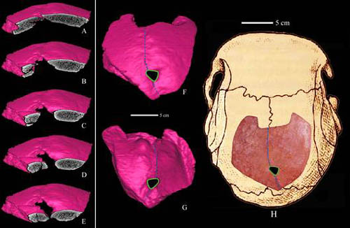
Fig.2. CT reconstruction of the Xujiyao 11 parietal bones with sequential coronal slices through the perforation. The slices extend from the anterior edge of the opening (A) to close to the posterior margin (E). A 3D CT reconstruction of the specimen (H) is shown in external (F) and internal (G) views. (Image by WU Xiujie)