| Location: Home > Research > Research Progress |
| Fossil Evidence Supports Developmental Model for the Origin of the Jaw |
|
The invention of the jaw is the most profound and radical evolutionary step in the vertebrate history. However, the sequence leading to the evolutionary origin of the jaw is still unclear largely due to the absence of data on the ancestral, intermediate condition.
In an article published in Chinese Science Bulletin (vol.57,No.30), Drs. GAI Zhikun and ZHU Min from the Institute of Vertebrate Paleontology and Paleoanthropology (IVPP), Chinese Academy of Sciences, reviewed the origin of the vertebrate jaw based on the molecular, developmental and paleontological evidences, and found that 435-370-million-year-old jawless galeaspids from China and northern Vietnam display an intermediate condition in the establishment of diplorhiny and jaw in which the barrier to the forward growth of neuralcrest-derived craniofacial ectomesenchyme was removed.This study provides a terrific example of intersection between developmental biology-based model and fossil evidence, and suggests that galeaspids might be the closest jawless relatives of jawed vertebrates. Advances in developmental genetics have accumulated to propose the heterotopy theory of jaw evolution, i.e. the jaw evolved as a novelty through a heterotopic shift of mesenchyme-epithelial interaction. According to this theory, the disassociation of the nasohypophyseal complex and the development of the trabeculae are two fundamental prerequisites for the origin of the jaw, since the median position of the nasohypophyseal placode in cyclostome head development precludes the forward growth of the neural-crest-derived craniofacial ectomesenchyme. The disassociation of the nasohypophyseal complex is also regarded as a major event for the evolutionary origin of the diplorhiny (nasal sacs with individual externalized nostrils) at the agnathan-gnathostome transition. Galeaspids provide the earliest evidence for the clear separation of the olfactory organs from the hypophyseal duct in vertebrate phylogeny, a prerequisite condition in evolutionary developmental models for the origin of the jaw and diplorhiny. No fossil evidence indicates that the diplorhinic condition (nasal sacs with individual externalized nostrils) has been established before the invention of the jaw, but an intermediate condition, paired laterally positioned nasal sacs and independent hypophyseal organ appeared in jawless galeaspids. Using Synchrotron Radiation X-ray Tomography, researchers observed some derivative structures of the trabeculae (e.g. orbitonasal lamina, ethmoid plate) in 435–370-million-year-old jawless galeaspids, which provide new insights into the reorganization of the vertebrate head before the evolutionary origin of the jaw. These anatomical observations via new techniques highlight the possibility that galeaspids are, in many respects, a better proxy than osteostracans for reconstructing the pre-gnathostome condition of the rostral part of the braincase. The cranial anatomy of galeaspids reveals a number of derived characters uniquely shared with gnathostomes. This raises the potential possibility that galeaspids might be the closest jawless relatives of jawed vertebrates. This work was supported by the National Natural Science Foundation of China, the National Basic Research Program of China, the Knowledge Innovation Program of the Chinese Academy of Sciences, and the CAS/SAFEA International Partnership Program for Creative Research Teams. 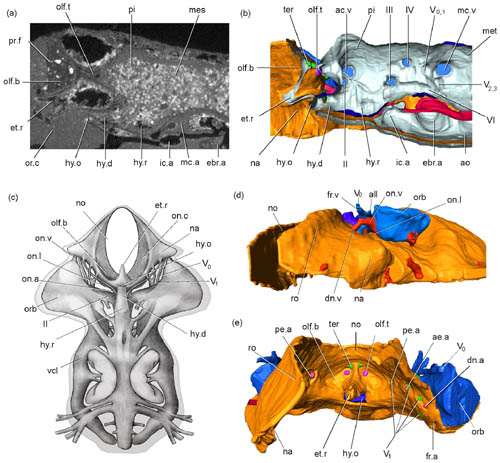 Fig.1 The cranial anatomy of Shuyu zhejiangensis, a 430-million-year-old jawless fish from Zhejiang, China. The derivative structures of the trabeculae (rostrum, orbitonasal lamina, ethmoid plate) and the relative nervous and vascular canals such as the superficial ophthalmic nerve, profundus nerve, orbitonasal artery, and orbitonasal vein are shown by the synchrotron radiation X-ray tomography and restoration. (image by GAI Zhikun) |