| Location: Home > Research > Research Progress |
| Peking Man Differing From Modern Humans in Brain Asymmetry |
|
Paleoanthropologists studying the fossil endocasts of Australopithecus, Homo habilis, Homo erectus, Neanderthals, and Homo sapiens have reported that almost all brain endocasts display distinct cerebral asymmetry. Peking man’s endocasts are good examples of ancestral brains and are useful in studying human evolution. However, studies examining brain asymmetries in fossil hominids are usually limited to scoring of differences in hemisphere protrusion rostrally and caudally, or to comparing the width of the hemispheres. Dr. WU XiuJie and PAN Lei, one of her graduate students, Institute of Vertebrate Paleontology and Paleoanthropology (IVPP), Chinese Academy of Sciences, using 3D laser scanning, examined asymmetries of the hemisphere volumes and surface areas in the Zhoukoudain Homo erectus, well known as Peking man, dated to 0.4–0.8 Ma. They found that Peking man exhibited a greater variation than the modern humans in the relative sizes between the left and right hemispheres, and no differences in the two hemispheres in Peking man, while in the modern endocasts the left hemisphere was significantly greater than the right hemisphere, as reported in the latest issues of Chinese Science Bulletin 56 (21):2215-2220. Brain evolution is one of the most important aspects of human evolution. For fossil hominids, the soft brain tissue was removed during fossilization. As such, endocasts are the direct material for the study of brain evolution. An endocast is an impression taken from the inside of a cranium that reflects the external features of the brain anatomy in detail. Six nearly complete crania from Peking man were discovered at Zhoukoudian Locality 1 in the suburbs of Beijing since the official excavation in 1927. From these, six endocasts were reconstructed from the original cranium fossils from Zhoukoudian Homo erectus. Dr. Wu used a 3D laser surface technique to scan the Peking man’s endocasts in order to reconstruct the 3D brain images. Using these methods Wu was able to calculate absolute and relative volumes and surface areas of two hemispheres for this study. Compared with modern humans, Peking man’s brain casts have small brain size, low height and low position of the greatest breadth, flat frontal and parietal lobes, depressed Sylvian areas, strong posterior projection of the occipital lobes, anterior positioning of the cerebellar lobes relative to the occipital lobes, and relative simplicity of the meningeal vessels. The study shows that the absolute hemisphere volumes and surface areas exhibited no significant asymmetries in the Peking man or in modern specimens. However, the relative hemisphere volumes against surface areas differed between the two groups, suggesting that brain asymmetries originated from relative brain sizes rather than absolute brain volumes during human evolution. These anatomical changes are likely related to the origin of human brain lateralization. The anatomical structures of Peking man’s brain maybe differs from the modern human, suggesting that Peking man had no ability to communicate with each other in the form of language. During human evolution, brain anatomical asymmetries experienced marked changes, and certain human abilities including language, intelligence, and cognition are likely related to these asymmetrical changes. “Although it was suggested that Peking man might exhibit a left hemisphere larger than the right hemisphere, studies restricted to the traditional method of examination were not able to examine these parameters. Using a 3D laser surface technique, we were able to calculate the absolute and relative volumes and surface areas of the two hemispheres”, said Dr. WU, the lead author and research designer. This work was supported by the National Natural Science Foundation of China, the Knowledge Innovative Program of the Chinese Academy of Sciences and the International Cooperation Program of MST of China.
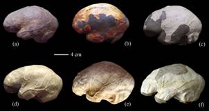
Fig.1: The six Peking man’s endocasts used in the study, showing left lateral views. (Images by WU XiuJie)
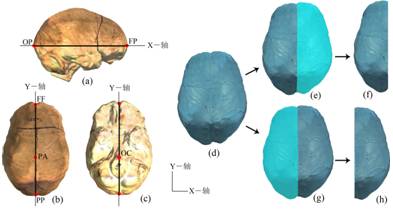
Fig.2: Virtual 3D endocasts of Peking man and methods used to split the left and right hemispheres. (a) Standard X-axis reference plane; (b), (c) standard Y-axis reference plane. (d)–(f) Methods used to split the left hemisphere; (d), (g), (h) methods used to split the right hemisphere. (Images by WU XiuJie) |